| | |
 | | DIgestive Tract Model Chart (9) | by GreenFlames09 on Flickr |
| 1. The Nose- - Draws air in through the nostrils to the pharynx.
2. Pharynx- - The throat.
- Connects the nose to the larynx.
3. Larynx - Voice box
- connects pharynx to trachea.
4. Trachea - Windpipe
- Connects larynx to the bronchus.
|
 | | cartilage | by jetheriot on Flickr |
| 5. Bronchi- Main tubes run from trachea to bronchioles.
6. Bronchioles- Lead into lungs.
- Made of smooth muscle.
|
 | | Normal lung: Alveoli | by Pulmonary Pathology on Flickr |
| 7. Alveoli- Air sacs in the lungs.
- Where gas exchange takes place.
- Smallest part of the respiratory system.
|
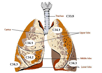 | | lung illustration | by Joe Crawford (artlung) on Flickr |
| Lungs- The largest, most important organs in respiration and ventilation.
- The right lung has three lobes and the left lung only has two.
- The heart tucks in between the two lungs and into the left lung.
- The diaphragm (not pictured), is a structure located beneath the lungs. The major function of the diaphragm is assisting with breathing.
- When breathing, the diaphragm will move. During inspiration, the diaphragm moves downward and the lungs expand, filling with oxygen. During expiration, the diaphragm moves upward to a normal position, and carbon dioxide is forced out of the lungs.
|
 NeoK12.com - Educational Videos, Lessons, Quizzes & Presentations