| Share Presentation: https://NeoK12.com/pres/ZCIRSYS1 | |
|
| ||||
|
Oxygenated blood travels from the PULMONARY VEIN to the left atrium, then onto the left ventricle and with deep contraction is pumped under high pressure through the aorta. This is THE ONLY VEIN IN THE BODY THAT CARRIES OXYGENATED BLOOD.
De-oxygenated blood (high in CO2 from cellular respiration) enters the right atrium from the inferior or superior vena cava to be then sent back to the lungs via the PULMONARY ARTERY. This is THE ONLY ARTERY IN THE BODY THAT CARRIES DEOXYGENATED BLOOD!
Running through the middle of the heart is a septum which separates the chambers. | ||||
|
The heart contains four valves. The valves are designed to prevent backflow of blood.
mitral valve - L atrium & L ventricle.
Aortic valve - L ventricle & Aorta
Pulmonic - R atrium & R ventricle
Tricuspid - R atrium & pulmonary artery lub dub - sound of the heart through a stethoscope
lub = closing of mitral & tricuspid valves dub = closing of aortic & pulmonary valves
YOUR HEART BEATS 60-80BPM AT REST! | ||||
|
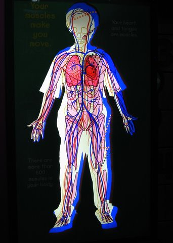
BLOOD VESSELS
Systemic arteries - thick, muscular, elastic walls that carry oxygenated blood under high pressure, from the heart to the rest of the body.
Systemic veins - thin walls, contain valves (to prevent backflow of CO2 rich blood) that take de-oxygenated blood from the bodies cells after cellular respiration back to the heart (which is then sent onto the lungs by the pulmonary artery for excretion - exhale)
Capillaries - one cell thick - allow the blood to exchange gases, nutrients and wastes with tissues. | ||||
|
BLOOD COMPONENTS
Red Blood Cells (RBC) = carry O2. Assist in transfusions if pt anaemic (& not requiring blood volume
Plasma = mostly water = increases blood volume. Assists in trauma when pt bleeding lots.
Platelets = clotting. Assists in pts with inability to clot effectively. E.g. haemophilia
White Blood Cells (WBC) = infection fighting cells. Assist in pt with decreased immunity and unable to fight infection with antibiotics. | ||||
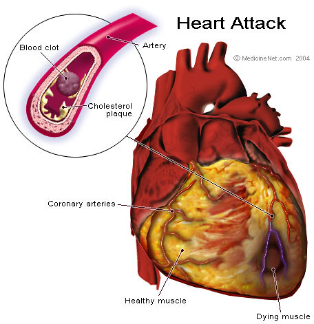
|
Any disease of the heart, blood or its vessels. Disease interrupts the flow of O2 to body organs CVD = Not Contagious CVD = Poor Lifestyle Choices (smoking, poor diet, high BP, lack of exercise) CVD = Signs/Symptoms...chest pain/discomfort, shortness of breath, sweating, nausea, fatigue. CVD - CardioVascular Disease | ||||